Cartilage Restoration
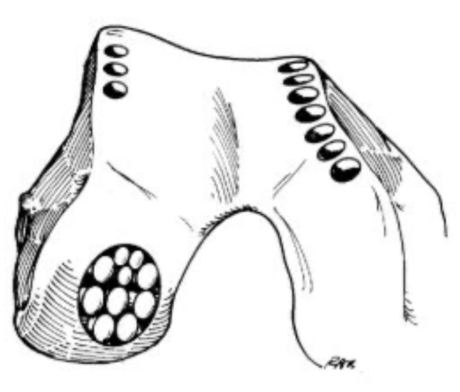
Biological resurfacing techniques including OATS, osteochondral allograft and autologous chondrocyte implantation for focal cartilage defects.
Overview
Cartilage restoration aims to regenerate or replace damaged articular cartilage in the knee, particularly in younger patients with focal defects. The goal is to prevent progression to widespread osteoarthritis and delay or avoid joint replacement surgery. Due to the poor longevity of structural repair and inferior tissue quality, microfracture is rarely performed in Mr Getgood's practice. Instead, the focus is on techniques that provide more durable, hyaline-like cartilage restoration.
Indications
- Focal full-thickness cartilage defects
- Osteochondritis dissecans lesions
- Traumatic cartilage injuries in young active patients
- Symptomatic cartilage defects on MRI
Recovery
6–12 months depending on technique.
Cartilage Restoration Procedures
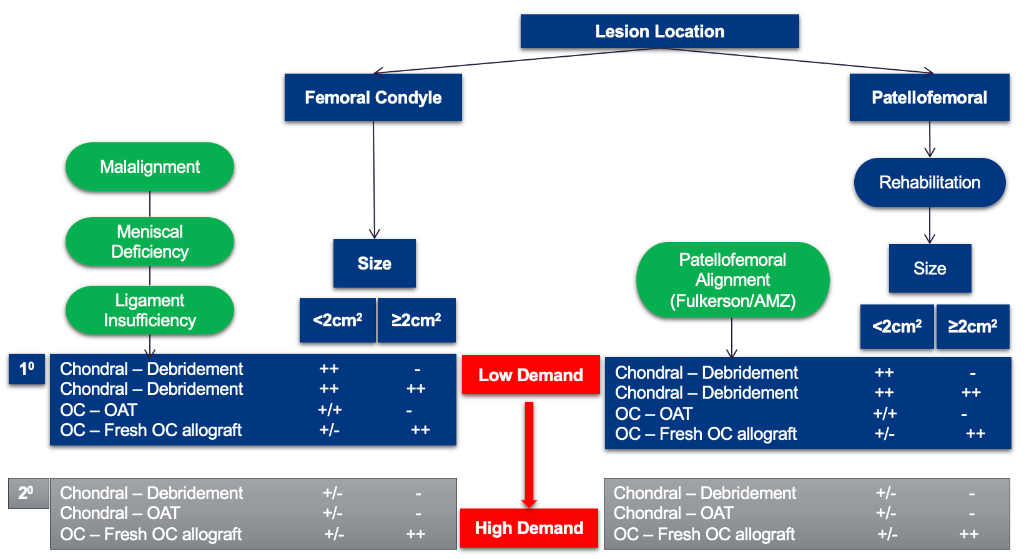
The choice of cartilage procedure depends on lesion size, location, patient demand, and prior treatment history. Mr Getgood will determine the most appropriate technique at consultation based on your MRI and clinical assessment.
Cartilage Restoration Decision Algorithm
The following algorithm guides treatment selection based on lesion location, size, and patient demand level.
Rehabilitation Programme
The goal of surgery is to relieve pain and restore the surface and congruity of the knee joint. This is not a procedure that aims to return patients to high-intensity, pivoting or impact sport. It is common for other procedures to be performed simultaneously — such as ACL reconstruction, meniscal allograft transplantation or realignment osteotomy — and rehabilitation will be tailored accordingly.
| Phase / Time | Brace & Weight Bearing | ROM | Exercises & Modalities |
|---|---|---|---|
Phase I Weeks 0–2 Protection & ROM | No brace unless specific instruction. Flat foot feather touch weight bearing with crutches. | Full range as tolerated. |
Modalities: Ice compression therapy and IFC as needed to reduce pain and swelling. Compex/muscle stimulation to assist quadriceps activation. Blood flow restriction (BFR) therapy during strengthening exercises. |
Phase II Weeks 2–8 Early Weight Bearing | No brace unless specific instruction. Partial weight bearing with crutches, encouraging normal heel–toe gait. | Full range as tolerated. |
Modalities: Ice compression therapy and IFC as needed to reduce pain and swelling. Compex/muscle stimulation to assist muscle activation. Blood flow restriction (BFR) therapy during strengthening exercises. |
Phase III Weeks 8–16 Weight Bearing & Strengthening | Advance to weight bear as tolerated. Wean off crutches, ensuring normal heel–toe gait. | Full range as tolerated. |
Modalities: Ice compression therapy as needed. Compex/muscle stimulation to assist muscle activation. Blood flow restriction (BFR) therapy during strengthening exercises. |
Phase IV 4–6 Months Strength & Conditioning | As tolerated per surgeon guidance. | Full and pain-free. |
Modalities: Blood flow restriction (BFR) therapy during strengthening exercises. |
Phase V 6+ Months Return to Activity / Light Sport | As tolerated per surgeon guidance. | Full and pain-free. | Impact activities such as jogging/running may commence from 6 months onwards. Note: long-distance running is not recommended.
|
Phase I — Weeks 0–2
Protection & ROM
Brace & Weight Bearing
No brace unless specific instruction. Flat foot feather touch weight bearing with crutches.
ROM
Full range as tolerated.
Exercises
- Patellar mobilisations
- Heel slides (± slider board), calf & hamstring stretches
- Quad/ham co-contraction, gluteal activation (supine or standing), isometric hip adduction/abduction, ankle pumping
- Sitting passive leg extension with roll under heel if extension lag
Modalities: Ice compression therapy and IFC as needed to reduce pain and swelling. Compex/muscle stimulation to assist quadriceps activation. Blood flow restriction (BFR) therapy during strengthening exercises.
Phase II — Weeks 2–8
Early Weight Bearing
Brace & Weight Bearing
No brace unless specific instruction. Partial weight bearing with crutches, encouraging normal heel–toe gait.
ROM
Full range as tolerated.
Exercises
- Patellar mobilisations
- Supine legs up wall: heel slides (knee flexion); supine legs on Swiss ball: knee flexion
- Bike pendulums: ½ circles forward/backward — lower seat as tolerated
- Progress to full revolutions, using contralateral limb to power cycle (home-based continuous passive motion)
- Prone hangs for extension if needed
- Quadriceps isometrics in long sitting and standing (± muscle stimulation)
- Non-weight bearing hip stability: abduction, extension, external rotation, clam shells, supine bridging on Swiss ball
- Ankle theraband plantar flexion, sitting calf raises
Modalities: Ice compression therapy and IFC as needed to reduce pain and swelling. Compex/muscle stimulation to assist muscle activation. Blood flow restriction (BFR) therapy during strengthening exercises.
Phase III — Weeks 8–16
Weight Bearing & Strengthening
Brace & Weight Bearing
Advance to weight bear as tolerated. Wean off crutches, ensuring normal heel–toe gait.
ROM
Full range as tolerated.
Exercises
- Continue patellar mobilisations
- Low resistance stationary bike
- Weight shifting: 2 scales 50–50 WB → progress to mini squats
- Leg extension (or quad over roll), active terminal knee extension with theraband
- Abdominal and core strengthening (curl-ups, transversus abdominis with SLR)
- Standing hip flexion/extension, abduction/adduction with weights/pulleys/bands
- Supine bridging: 2 → 1 leg Swiss ball bridge + knee flexion
- Shuttle/leg press: 2-leg squat/calf raises → progress 2 → 1 leg; increase ROM & resistance
- Mini wall squats (30–60°) → 60–90°; sit to stand
- Hamstring curls: prone and sitting → progress 1–2 lb weights
- Calf raises 2 → 1 foot; up-on-toes walking (when full WB)
- Wobble boards with support: side-to-side, forward/backward
- Single leg stance 30–60 seconds (when full WB)
Modalities: Ice compression therapy as needed. Compex/muscle stimulation to assist muscle activation. Blood flow restriction (BFR) therapy during strengthening exercises.
Phase IV — 4–6 Months
Strength & Conditioning
Brace & Weight Bearing
As tolerated per surgeon guidance.
ROM
Full and pain-free.
Exercises
- Continue bike; add elliptical
- Stairmaster if adequate strength (no hip hiking when pressing down on step)
- Continue core strengthening (obliques, planks, Pilates)
- Progress leg extensions with weight as tolerated (pain-free arc)
- Sit to stand lower seat height → single leg (watch mechanics)
- Progress resistance on shuttle: strength & endurance, 2 → 1 leg
- Static lunge → dynamic lunge → lunge walking (within pain-free range)
- Bungee cord walking: forward, backward, side step, lunging → add speed/direction change
- Forward and lateral step-ups 2–4–6" and eccentric lateral step-down with control
- Squats and lunges on Dynadisc / Airex / BOSU (within range as tolerated)
- Continue hip strengthening: weights, pulleys, tubing; tubing kick-backs (mule kicks)
- Shuttle standing kick-backs (hip/knee extension)
- Supine Swiss ball bridge + knee flexion → 1 leg
- Chair walking / stool pulls
- Hamstring curls: standing & sitting — weights/pulleys/bungee; eccentric heel drops 2 → 1 leg
- Continue wobble boards; add upper body skills (throwing, catching)
- Single leg stance on unstable surfaces (pillow, mini-tramp, BOSU, Airex, Dynadisc)
- Single leg stance with upper body patterning specific to patient goals
- Standing 747s: eyes open/closed → progress to mini trampoline
Modalities: Blood flow restriction (BFR) therapy during strengthening exercises.
Phase V — 6+ Months
Return to Activity / Light Sport
Brace & Weight Bearing
As tolerated per surgeon guidance.
ROM
Full and pain-free.
Exercises
Impact activities such as jogging/running may commence from 6 months onwards. Note: long-distance running is not recommended.
- Begin jogging/running programme once full ROM regained with quiet knee and adequate neuromuscular control
- Agility: cariocas/grapevine, figure 8s around cones, ladder drills, lateral shuffle cone-to-cone
- Side-to-side steps/jumps on BOSU
- Line jumping: backward/forward/side-to-side → progress to diagonals/combined patterns; 2 → 1 leg
- Jumping: tuck jumps, box jumps, long jumps
- Skipping rope: double and single leg
- Hopping: single-leg distance, 6 m timed, triple hop, cross-over — 2 → 1 leg
- Sport-specific multi-directional drills and contact when adequate core/lower extremity patterning (stop-and-go, sideways/backwards drills, sprinting with cutting and pivoting)
* Timelines are approximate. Exercises are given as examples only and should be directed by your physiotherapist. Progression is criteria-based and directed by your physiotherapist in conjunction with Mr Getgood's clinical review. Individual recovery varies.
Frequently Asked Questions
Supporting Evidence
Noteworthy Publications from Mr Getgood Pertaining to Cartilage Restoration
Whittaker JL, Kalsoum R, Bilzon J, Conaghan PG, Crossley K, Dodge GR, et al. Toward designing human intervention studies to prevent osteoarthritis after knee injury: A report from an interdisciplinary OARSI 2023 workshop. Osteoarthr Cartil Open. 2024;6(2):100449.
Nakanishi Y, Hegarty P, Vivacqua T, Firth A, Milner JS, Pritchett S, et al. Quantitative MRI Analysis of Patellofemoral Joint Cartilage Health 2 Years After Anterior Cruciate Ligament Reconstruction and Lateral Extra-Articular Tenodesis. Am J Sports Med. 2024;52(7):1773–83.
Firth AD, Pritchett SL, Milner JS, Atkinson HF, Bryant DM, Holdsworth DW, et al. Quantitative Magnetic Resonance Imaging of Lateral Compartment Articular Cartilage After Lateral Extra-articular Tenodesis. Am J Sports Med. 2024;52(4):909–18.
Gortz S, Tabbaa SM, Jones DG, Polousky JD, Crawford DC, Committee M, et al. Metrics of OsteoChondral Allografts (MOCA) Group Consensus Statements on the Use of Viable Osteochondral Allograft. Orthop J Sports Med. 2021;9(3):2325967120983604.
Chahla J, Hinckel BB, Yanke AB, Farr J, Metrics of Osteochondral Allografts G, Bugbee WD, et al. An Expert Consensus Statement on the Management of Large Chondral and Osteochondral Defects in the Patellofemoral Joint. Orthop J Sports Med. 2020;8(3):2325967120907343.
Other Noteworthy Publications Pertaining to Cartilage Restoration
de Girolamo L, Filardo G, Abat F, Barfod KW, Bastos R, Cugat R, et al. The use of injectable orthobiologics for knee osteoarthritis: A formal ESSKA-ORBIT consensus. Part 2-Cell-based therapy. Knee Surg Sports Traumatol Arthrosc. 2025;33(11):4079–95.
Diaz Haaz DI, Rizo Castro O. Efficacy and Safety of Intra-articular Platelet-Rich Plasma (PRP) Versus Corticosteroid Injections in the Treatment of Knee Osteoarthritis: A Systematic Review of Randomized Clinical Trials. Cureus. 2025;17(3):e80948.
Kjennvold S, Rotterud JH, Forssblad M, Engebretsen L, Aroen A, Ulstein S. Debridement or Microfracture for Full-Thickness Cartilage Lesions in Anterior Cruciate Ligament Reconstruction: A 10-Year Cohort Study of 326 Patients in Norway and Sweden. Orthop J Sports Med. 2025;13(10):23259671251381340.
Laver L, Filardo G, Sanchez M, Magalon J, Tischer T, Abat F, et al. The use of injectable orthobiologics for knee osteoarthritis: A European ESSKA-ORBIT consensus. Part 1-Blood-derived products (platelet-rich plasma). Knee Surg Sports Traumatol Arthrosc. 2024;32(4):783–97.
Rotterud JH, Sivertsen EA, Forssblad M, Engebretsen L, Aroen A. Effect on Patient-Reported Outcomes of Debridement or Microfracture of Concomitant Full-Thickness Cartilage Lesions in Anterior Cruciate Ligament-Reconstructed Knees: A Nationwide Cohort Study From Norway and Sweden of 357 Patients With 2-Year Follow-up. Am J Sports Med. 2016;44(2):337–44.