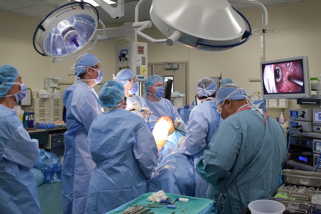
Knee Arthroscopy
Minimally invasive surgical procedures for diagnostic and therapeutic treatment of knee joint problems
Overview
Knee arthroscopy is a minimally invasive surgical technique using a small camera (arthroscope) to visualize and treat various knee conditions. Common procedures performed through arthroscopy include: • Partial Meniscectomy: Removal of torn portions of the meniscus to relieve pain and improve joint function • Loose Body Removal: Extraction of loose bone or cartilage fragments that may be causing mechanical symptoms • Chondroplasty: Smoothing of damaged or degraded cartilage surfaces to improve joint mechanics These arthroscopic procedures are among the most common and safest knee interventions, with minimal tissue damage, rapid recovery, and excellent safety profiles.
Indications
- Torn meniscus causing mechanical symptoms
- Loose bodies within the joint
- Cartilage damage or chondral lesions
- Synovitis or joint inflammation
- Ligamentous injuries requiring evaluation
Recovery
2-4 weeks for return to light activities; 10-16 weeks for full return to sport and activities
Operative vs. Non-operative Treatment
There is a lot of discussion in the media and medical literature regarding the lack of evidence for knee arthroscopy. A number of clinical trials have shown that physical therapy will often have a similar treatment effect to knee arthroscopy for the treatment of degenerative meniscus tears (see supporting evidence section). There are clear indications where knee arthroscopy remains very effective (see above). In the case of degenerative tears, Mr Getgood will advise on the best course of initial management, utilising physical therapy and injection treatment with the aim of avoiding operative intervention.
Rehabilitation Programme
Recovery following knee arthroscopy is typically faster than major reconstructive surgery, but the joint still requires a structured, progressive rehabilitation programme to ensure full restoration of strength, range of motion and function. All progressions are criteria-based and directed by your physiotherapist in conjunction with Mr Getgood's post-operative instructions.
| Phase / Time | Goals | Suggested Exercises and Guidelines |
|---|---|---|
Phase I Weeks 0–2 Acute Recovery | Control pain and swelling, restore full extension, begin range of motion. | NO plyometric or impact activities. No running. Protect the joint.
|
Phase II Weeks 2–4 Early Strengthening | Achieve full range of motion, wean off crutches, begin closed-chain strengthening. | NO plyometric, jumping or running activities in the first 4 weeks. The joint requires this time to recover from surgical trauma and reduce intra-articular inflammation.
|
Phase III Weeks 4–8 Progressive Loading | Build lower limb strength and neuromuscular control, begin sport-specific conditioning. |
|
Phase IV Weeks 8–12+ Return to Sport / Activity | Full return to sport, recreational activity or work demands. Achieve limb symmetry. |
|
Phase I — Weeks 0–2
Acute Recovery
Goals
Control pain and swelling, restore full extension, begin range of motion.
Exercises & Guidelines
NO plyometric or impact activities. No running. Protect the joint.
- Rest, ice (20 min every 2 hours), compression and elevation (RICE)
- Crutches as required — weight bear as tolerated
- Ankle pumps and calf stretches to reduce DVT risk
- Quadriceps sets (isometric): 3 × 10 reps
- Heel slides for knee flexion — progress as tolerated
- Straight leg raises (SLR): 3 × 10 reps
- Prone hangs for full extension if extension lag present
- Patella mobilisations: medial, inferior and superior glides
- Ice and TENS/IFC for analgesia as needed
- Compex/muscle stimulation to assist quadriceps activation
- Blood flow restriction (BFR) therapy during lower limb exercises
Phase II — Weeks 2–4
Early Strengthening
Goals
Achieve full range of motion, wean off crutches, begin closed-chain strengthening.
Exercises & Guidelines
NO plyometric, jumping or running activities in the first 4 weeks. The joint requires this time to recover from surgical trauma and reduce intra-articular inflammation.
- Wean off crutches once walking with a normal gait pattern
- Stationary bike: low resistance, seat raised (begin when flexion ≥ 100°)
- Mini squats: 0–60° — 3 × 15 reps
- Sit to stand from a raised chair — progress seat height down
- Terminal knee extension with theraband
- Standing hip abduction, extension and flexion with theraband
- Calf raises: 2-leg → 1-leg progression
- Aquatic physiotherapy if wound healed (from Week 3)
- Hydrotherapy walking and range of motion drills
- Ice compression therapy to manage swelling and pain
- Compex/muscle stimulation to assist muscle activation
- Blood flow restriction (BFR) therapy during strengthening exercises
Phase III — Weeks 4–8
Progressive Loading
Goals
Build lower limb strength and neuromuscular control, begin sport-specific conditioning.
Exercises & Guidelines
- Progress squats to 90°: 2-leg → single-leg (monitor mechanics)
- Leg press: bilateral → unilateral, increase resistance progressively
- Lateral step-ups and eccentric step-downs: 4–6 inch step
- Forward and reverse lunges
- Romanian deadlift for hamstring strength
- Single-leg stance on stable → unstable surfaces (wobble board, Airex)
- Begin jogging programme when single-leg squat control is adequate and effusion is minimal
- Elliptical trainer and Stairmaster
- Core stability: planks, bridges, pallof press
- Ice compression therapy as needed for swelling management
- Compex/muscle stimulation to assist muscle activation
- Blood flow restriction (BFR) therapy during strengthening exercises
Phase IV — Weeks 8–12+
Return to Sport / Activity
Goals
Full return to sport, recreational activity or work demands. Achieve limb symmetry.
Exercises & Guidelines
- Running programme: progressive distance and speed
- Agility drills: cariocas, lateral shuffles, figure-of-8, ladder drills
- Plyometrics: introduce double-leg jumping, box jumps, bounding (when strength symmetry ≥ 80% LSI)
- Sport-specific cutting, pivoting and directional change drills
- Single-leg hop testing: single, triple and crossover hops
- Return to full training and competition when cleared by surgeon and physiotherapist
- Ongoing strength maintenance programme
- Blood flow restriction (BFR) therapy during strengthening exercises (continue for first 6 months)
* Timelines are approximate. Exercises are given as examples only and should be directed by your physiotherapist. Progression is criteria-based and directed by your physiotherapist in conjunction with Mr Getgood's clinical review. Individual recovery varies depending on the procedure performed arthroscopically.
Frequently Asked Questions
Supporting Evidence
Noteworthy Publications from Mr Getgood Pertaining to Knee Arthroscopy and the Treatment of Early Osteoarthritis
Arthroscopy Association of Canada, Wong I, Hiemstra L, Ayeni OR, Getgood A, Beavis C, et al. Position Statement of the Arthroscopy Association of Canada (AAC) Concerning Arthroscopy of the Knee Joint—September 2017. Orthop J Sports Med. 2018.
Chahla J, Hinckel BB, Yanke AB, Farr J, Metrics of Osteochondral Allografts G, Bugbee WD, et al. An Expert Consensus Statement on the Management of Large Chondral and Osteochondral Defects in the Patellofemoral Joint. Orthop J Sports Med. 2020.
Degen RM, Lebedeva Y, Birmingham TB, Marsh JD, Getgood AMJ, Giffin JR, et al. Trends in knee arthroscopy utilization: a gap in knowledge translation. Knee Surg Sports Traumatol Arthrosc. 2020.
Malone A, Price J, Price N, Peck V, Getgood A, Petrella R, et al. Safety and pharmacokinetics of EP-104IAR (sustained-release fluticasone propionate) in knee osteoarthritis: A randomized, double-blind, placebo-controlled phase 1 trial. Osteoarthr Cartil Open. 2021.
Thornton J, Barton KI, Churchill L, Schulz JM, Bryant D, Ambrose A, et al. Novel uses of healthcare technology for individuals with mild to moderate hip or knee osteoarthritis: The technology, exercise and activity prescription for enhanced mobility (TEAM) study randomized controlled trial protocol. Osteoarthr Cartil Open. 2025.
Other Noteworthy Publications Pertaining to Knee Arthroscopy
Katz JN, Brophy RH, Chaisson CE, de Chaves L, Cole BJ, Dahm DL, et al. Surgery versus physical therapy for a meniscal tear and osteoarthritis. N Engl J Med. 2013;368(18):1675–84.
Kirkley A, Birmingham TB, Litchfield RB, Giffin JR, Willits KR, Wong CJ, et al. A randomized trial of arthroscopic surgery for osteoarthritis of the knee. N Engl J Med. 2008;359(11):1097–107.
Sihvonen R, Paavola M, Malmivaara A, Itala A, Joukainen A, Nurmi H, et al. Arthroscopic partial meniscectomy versus sham surgery for a degenerative meniscal tear. N Engl J Med. 2013;369(26):2515–24.