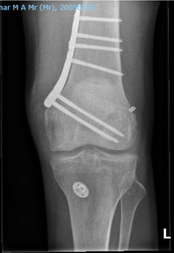
Knee Osteotomy
Realignment surgery correcting varus or valgus deformity to redistribute load away from the damaged compartment, delaying the need for joint replacement.
Overview
High tibial osteotomy (HTO) or distal femoral osteotomy (DFO) are joint-preserving procedures that correct mechanical axis malalignment. By realigning the limb, load is transferred from the worn compartment to healthy cartilage. The procedure involves a controlled bone cut with gradual opening or closing of the osteotomy site, fixed with a plate and screws. Modern planning uses long-leg weight-bearing radiographs and sometimes 3D-printed patient-specific guides for precision. Osteotomy is particularly valuable in young, active patients with unicompartmental arthritis who are not ideal candidates for joint replacement.
Indications
- Unicompartmental osteoarthritis in young patients
- Malalignment causing asymmetric joint wear
- Medial compartment overload with varus deformity
- Lateral compartment overload with valgus deformity
Recovery
3–4 months for bone healing; 6 months for full activity.
Patient Information Video
Mr Getgood works with an Australian company called Personalised Surgery that has produced the following patient information video on knee osteotomy.
Types of Osteotomy
The specific osteotomy technique is selected based on the location and magnitude of deformity, the compartment affected, and any associated pathology. Mr Getgood will determine the most appropriate procedure at consultation using your full-length standing X-rays, 3D CT scan and clinical assessment.
Rehabilitation Programme
The goal of realignment osteotomy surgery is to reduce the load placed through a specific part of the knee, thereby reducing pain and protecting the ligaments or articular cartilage. It is common for other procedures to be performed simultaneously — such as ACL reconstruction, meniscus transplant, or cartilage repair — and rehabilitation will be tailored accordingly.
A similar programme is followed for both femoral (DFO) and tibial (HTO) osteotomies, whether opening or closing wedge, when using internal locking plate fixators.
| Phase / Time | Brace & Weight Bearing | ROM | Exercises & Modalities |
|---|---|---|---|
Phase I Weeks 0–2 Immediate Post-op | Tracker brace locked only when ambulating; full ROM allowed during exercises; worn 24 hours per day (can be removed during therapy). Flat foot feather touch weight bearing with crutches. | Full range of motion. |
Modalities: Ice compression therapy and IFC as needed to reduce pain and swelling. Compex/muscle stimulation to assist quadriceps activation. Blood flow restriction (BFR) therapy during strengthening exercises. |
Phase II Weeks 2–6 Progressive WB & Strengthening | Advance to weight bearing as tolerated with crutches; continue tracker brace until adequate quad control. Ensure normal heel–toe gait. Can remove brace when sleeping. | Progress to full ROM as tolerated (flexion and extension). |
Modalities: Ice compression therapy and analgesics as needed to reduce pain and swelling. Compex/muscle stimulation to assist muscle activation. Blood flow restriction (BFR) therapy during strengthening exercises. |
Phase III Weeks 6–12 Advanced Strengthening | No brace. Full weight bearing. Ensure normal heel–toe gait. | Full and pain-free knee range of motion. |
Modalities: Ice compression therapy and analgesics as needed to reduce pain and swelling. Compex/muscle stimulation to assist muscle activation. Blood flow restriction (BFR) therapy during strengthening exercises. |
Phase IV 3–6+ Months Return to Activity / Light Sport | As tolerated per surgeon guidance. | Full and pain-free. |
Modalities: Blood flow restriction (BFR) therapy during strengthening exercises. |
Phase I — Weeks 0–2
Immediate Post-op
Brace & Weight Bearing
Tracker brace locked only when ambulating; full ROM allowed during exercises; worn 24 hours per day (can be removed during therapy). Flat foot feather touch weight bearing with crutches.
ROM
Full range of motion.
Exercises
- Patellar mobilisations
- Heel slides (± slider board), calf & hamstring stretches
- Quad/ham co-contraction, gluteal activation (supine or standing), isometric hip adduction/abduction, ankle pumping
- Sitting passive leg extension with roll under heel if extension lag
- Non-weight bearing hip stability: abduction, extension, external rotation, clam shells, supine bridging on Swiss ball
- Ankle theraband plantar flexion, sitting calf raises
Modalities: Ice compression therapy and IFC as needed to reduce pain and swelling. Compex/muscle stimulation to assist quadriceps activation. Blood flow restriction (BFR) therapy during strengthening exercises.
Phase II — Weeks 2–6
Progressive WB & Strengthening
Brace & Weight Bearing
Advance to weight bearing as tolerated with crutches; continue tracker brace until adequate quad control. Ensure normal heel–toe gait. Can remove brace when sleeping.
ROM
Progress to full ROM as tolerated (flexion and extension).
Exercises
- Patellar mobilisations
- Sitting passive leg extension with roll under heel
- Supine with legs up wall — heel slides (knee flexion) gravity-assisted
- Bike pendulums: high seat ½ circles forward/backward → full circles — lower seat as tolerated
- Progress 2 crutches → single crutch → full WB, maintaining normal walking pattern
- Mini wall squat (30°) → progress to 60–90° (± wall)
- Shuttle (one bungee cord): 2-leg squat (¼–½ range) and 2-leg calf raises → progress slowly to 1-leg, increasing ROM & resistance
- Sit to stand 2 legs with high seat height (± muscle stimulation)
- Supine on floor, legs on Swiss ball: bridging → bridging + knee flexion (heels to buttocks)
- Wobble boards with support through full ROM: side-to-side, forward/backward
Modalities: Ice compression therapy and analgesics as needed to reduce pain and swelling. Compex/muscle stimulation to assist muscle activation. Blood flow restriction (BFR) therapy during strengthening exercises.
Phase III — Weeks 6–12
Advanced Strengthening
Brace & Weight Bearing
No brace. Full weight bearing. Ensure normal heel–toe gait.
ROM
Full and pain-free knee range of motion.
Exercises
- Sit to stand 2 legs → progress by decreasing seat height
- Bungee cord walking: forward / backward / side step with slow controlled return
- Static lunge (¼–½ range) → dynamic lunge step (¼–½ range) with proper alignment (shoulders over knees over toes)
- Step ups and down 2–4" height: lateral and forward
- Wobble boards side-to-side, forward/backward: without support, without vision
- Single leg stance 30–60 seconds (when full WB) → progress to unstable surface with and without vision
- Cardiovascular fitness conditioning
Modalities: Ice compression therapy and analgesics as needed to reduce pain and swelling. Compex/muscle stimulation to assist muscle activation. Blood flow restriction (BFR) therapy during strengthening exercises.
Phase IV — 3–6+ Months
Return to Activity / Light Sport
Brace & Weight Bearing
As tolerated per surgeon guidance.
ROM
Full and pain-free.
Exercises
- Progress resistance on shuttle: strength & endurance, 2 → 1 leg
- Static lunge (full range) → dynamic lunge → lunge walking
- Forward and lateral step-ups 4–6–8" (watch for hip hiking or excessive ankle dorsiflexion)
- Eccentric lateral step-down on 2–4–6" step with control
- Shuttle standing kick-backs (hip/knee extension)
- Single leg stance on unstable surfaces (pillow, mini-tramp, BOSU, Airex, Dynadisc) with/without support → progress to no vision
- Cardiovascular fitness conditioning
- Sport-specific training as indicated
Modalities: Blood flow restriction (BFR) therapy during strengthening exercises.
* Timelines are approximate. Exercises are given as examples only and should be directed by your physiotherapist. Progression is criteria-based and directed by your physiotherapist in conjunction with Mr Getgood's clinical review. Individual recovery varies.
Frequently Asked Questions
Supporting Evidence
Noteworthy Publications from Mr Getgood Pertaining to Osteotomy
Maniar AR, Mackay ND, Somerville L, Litchfield R, Lanting BA, Getgood AMJ. Improved clinical outcomes when maintaining a neutral joint line obliquity at high tibial osteotomy and subsequent total knee arthroplasty. J Exp Orthop. 2026;13(1):e70612.
Wackerle AM, Marcaccio S, Apseloff N, Getgood A, Musahl V, Tapasvi S, et al. Assessment and surgical correction of posterior tibial slope in revision anterior cruciate ligament surgery: An international expert Delphi consensus statement. J ISAKOS. 2025;13:100900.
Fritz J, Getgood A, van Heerwaarden R, Parratte S, Brown C, Tollefson LV, et al. Combined revision ACL reconstruction with slope-correction osteotomy and lateral extra-articular tenodesis improves stability in patients with high posterior tibial slope and pivot shift. J Exp Orthop. 2025;12(3):e70384.
Birmingham TB, Primeau CA, Moyer RF, Bryant DM, Ma J, Leitch KM, et al. High Tibial Osteotomy for Medial Compartment Knee Osteoarthritis: A Randomized Trial With Parallel Preference Arm. Ann Intern Med. 2025;178(9):1238–48.
Vivacqua T, Thomassen S, Winkler PW, Lucidi GA, Rousseau-Saine A, Firth AD, et al. Closing-Wedge Posterior Tibial Slope-Reducing Osteotomy in Complex Revision ACL Reconstruction. Orthop J Sports Med. 2023;11(1):23259671221144786.
Sidhu R, Moatshe G, Firth A, Litchfield R, Getgood A. Low rates of serious complications but high rates of hardware removal after high tibial osteotomy with Tomofix locking plate. Knee Surg Sports Traumatol Arthrosc. 2021;29(10):3361–7.
Cotter EJ, Gowd AK, Bohl DD, Getgood A, Cole BJ, Frank RM. Medical Comorbidities and Functional Dependent Living Are Independent Risk Factors for Short-Term Complications Following Osteotomy Procedures about the Knee. Cartilage. 2020;11(4):423–30.
Getgood A, Collins B, Slynarski K, Kurowska E, Parker D, Engebretsen L, et al. Short-term safety and efficacy of a novel high tibial osteotomy system: a case controlled study. Knee Surg Sports Traumatol Arthrosc. 2013;21(1):260–9.
Collins B, Getgood A, Alomar AZ, Giffin JR, Willits K, Fowler PJ, et al. A case series of lateral opening wedge high tibial osteotomy for valgus malalignment. Knee Surg Sports Traumatol Arthrosc. 2013;21(1):152–60.
Other Noteworthy Publications Pertaining to Osteotomy
Patel R, Mabrouk A, Paul S, Jacquet C, Ollivier M. Infra-tubercle osteotomy preserves coronal alignment and reduces anterior laxity compared to retro-tubercle technique in revision anterior cruciate ligament reconstruction with slope correction. Knee Surg Sports Traumatol Arthrosc. 2026;34(3):1024–34.
Hiranaka T, Kulkarni S, Grasso S, Parker DA. Accurate and reproducible coronal and sagittal alignment correction using patient-specific instrumentation in medial opening wedge high tibial osteotomy. J ISAKOS. 2026;17:101091.
Murphy T, Brady G, Dan M, Ollivier M, Caldwell B, Ghandour M. The ALPS classification: Predicting ACL graft failure based on deviation from mean posterior tibial slope—A systematic review and meta-analysis. Knee Surg Sports Traumatol Arthrosc. 2025.
Hiranaka T, Davey C, Grasso S, Dal Fabbro G, Ahedi H, Fritsch B, et al. A validated method accounting for joint line convergence angle reduces planning errors in medial opening wedge high tibial osteotomy. Knee Surg Sports Traumatol Arthrosc. 2025;33(9):3341–9.
Birmingham TB, Moyer R, Leitch K, Chesworth B, Bryant D, Willits K, et al. Changes in biomechanical risk factors for knee osteoarthritis and their association with 5-year clinically important improvement after limb realignment surgery. Osteoarthritis Cartilage. 2017;25(12):1999–2006.