Patellofemoral Stabilisation
Surgical correction for recurrent patellar dislocations using MPFL reconstruction and tibial tubercle osteotomy when indicated.
Overview
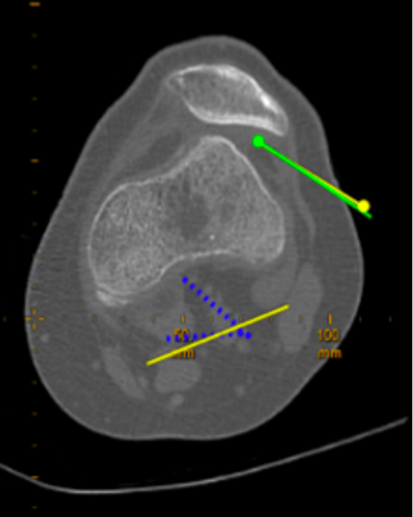
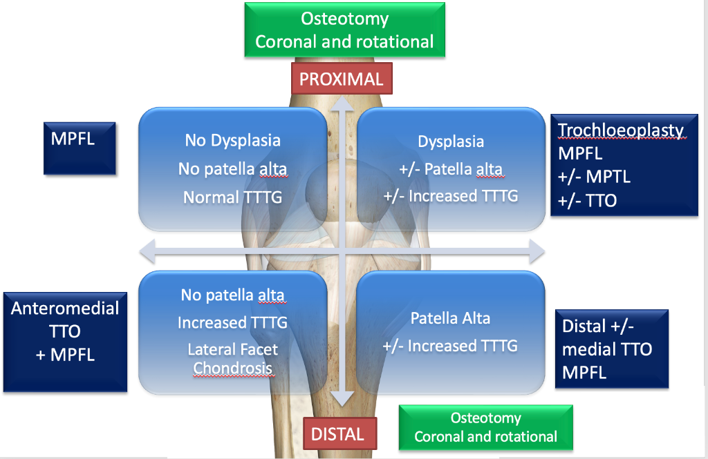
Patellofemoral instability surgery addresses recurrent dislocation or subluxation of the kneecap. The cornerstone procedure is medial patellofemoral ligament (MPFL) reconstruction, which restores the primary soft tissue restraint against lateral patellar displacement. In patients with underlying bony malalignment — such as an elevated tibial tubercle-trochlear groove distance (TT-TG) — a tibial tubercle osteotomy (TTO) may be combined with MPFL reconstruction to realign the extensor mechanism. Trochleoplasty may be considered in severe trochlear dysplasia to deepen the femoral groove and improve patellar tracking.
Indications
- Recurrent patellar dislocations
- First-time dislocation with osteochondral fragment
- Trochlear dysplasia with instability
- Failed conservative management
Recovery
4–6 months; return to sport at 6–9 months.
Stabilisation Procedures
Patellofemoral stabilisation is tailored to the underlying anatomical abnormality identified for each patient. Mr Getgood will determine the appropriate combination of procedures at consultation based on your clinical assessment, X-rays, and MRI or CT findings.
Patellofemoral Instability Surgery Algorithm
Rehabilitation Programme
Rehabilitation following patella stabilisation surgery is tailored to the specific technique(s) performed. Tibial tubercle osteotomy (TTO) requires a more protected early phase to allow bone healing, while isolated MPFL reconstruction and trochleoplasty have a faster initial progression. All progressions are criteria-based and directed by your physiotherapist in conjunction with Mr Getgood's clinical review.
| Phase / Time | Brace & Weight Bearing | ROM | Exercises |
|---|---|---|---|
Phase I Weeks 0–6 Protection & ROM | If TTO: brace locked in extension for ADLs, sleeping & ambulation (full WB in brace); remove for ROM/exercises (flat foot touch WB without brace). If no TTO: no brace, WBAT with crutches. Weeks 4–6: wean from 2→1 crutch with adequate quad control → full WB. | If TTO: 0–30° (weeks 0–2) → 0–60° (weeks 2–4) → 0–90° (weeks 4–6). If no TTO: increase as tolerated with steady weekly gains. | ⚠ TTO PATIENTS: NO active knee extension × 6 weeks
|
Phase II Weeks 6–12 Weight Bearing & Strengthening | TTO patients: advance to WBAT and wean off brace. All patients: wean off crutches. | All patients: progress to full ROM as tolerated (flexion and extension). |
|
Phase III 3–6+ Months Advanced Strengthening & Return to Activity | As tolerated per surgeon guidance. | Full range of motion. |
|
Phase I — Weeks 0–6
Protection & ROM
Brace & Weight Bearing
If TTO: brace locked in extension for ADLs, sleeping & ambulation (full WB in brace); remove for ROM/exercises (flat foot touch WB without brace). If no TTO: no brace, WBAT with crutches. Weeks 4–6: wean from 2→1 crutch with adequate quad control → full WB.
ROM
If TTO: 0–30° (weeks 0–2) → 0–60° (weeks 2–4) → 0–90° (weeks 4–6). If no TTO: increase as tolerated with steady weekly gains.
Exercises
⚠ TTO PATIENTS: NO active knee extension × 6 weeks
- Weeks 0–2: heel slides (± slider board), calf & hamstring stretches
- Quad/ham co-contraction, gluteal activation (supine or standing), isometric hip adduction/abduction, ankle pumping
- Sitting passive leg extension with roll under heel if extension lag
- Weeks 2–6 add: patellar mobilisations — medial, inferior & superior
- Supine legs up wall: heel slides; supine legs on Swiss ball: knee flexion
- Bike pendulums: ½ circles forward/backward → full circles (lower seat as tolerated)
- Prone hangs for extension if needed
- Quadriceps isometrics in long sitting and standing (± muscle stimulation)
- Non-WB hip stability: abduction, extension, external rotation, clam shells, supine bridging on Swiss ball
- Ankle theraband plantar flexion, sitting calf raises
- Ice and IFC as needed
- Ice compression therapy to manage swelling and pain
- Compex/muscle stimulation to assist quadriceps activation (± alongside isometrics)
- Blood flow restriction (BFR) therapy during lower limb exercises
Phase II — Weeks 6–12
Weight Bearing & Strengthening
Brace & Weight Bearing
TTO patients: advance to WBAT and wean off brace. All patients: wean off crutches.
ROM
All patients: progress to full ROM as tolerated (flexion and extension).
Exercises
- Continue patellar mobilisations: medial, inferior & superior
- Low resistance stationary bike
- Weight shifting: 2 scales 50–50 WB → progress to mini squats
- Leg extension / quad over roll, active terminal knee extension with theraband
- Abdominal and core strengthening (curl-ups, transversus abdominis with SLR)
- Standing hip flexion/extension, abduction/adduction with weights/bands
- Supine bridging: 2→1 leg Swiss ball bridge + knee flexion
- Shuttle / leg press: 2-leg squat/calf raises → 1-leg; increase ROM & resistance
- Mini wall squats 30–60° → 60–90°; sit to stand
- Hamstring curls: prone and sitting → progress 1–2 lb weights
- Calf raises 2→1 foot; up-on-toes walking (when full WB)
- Wobble boards with support: side-to-side, forward/backward
- Single leg stance 30–60 seconds (when full WB)
- Ice compression therapy to manage swelling and pain
- Compex/muscle stimulation to assist muscle activation
- Blood flow restriction (BFR) therapy during strengthening exercises
Phase III — 3–6+ Months
Advanced Strengthening & Return to Activity
Brace & Weight Bearing
As tolerated per surgeon guidance.
ROM
Full range of motion.
Exercises
- Continue bike; add elliptical and Stairmaster (if adequate strength — no hip hike)
- Continue core strengthening (obliques, planks, Pilates)
- Progress leg extensions with weight (pain-free arc)
- Sit to stand → single leg (watch mechanics)
- Progress resistance on shuttle: strength & endurance, 2→1 leg
- Static lunge → dynamic lunge → lunge walking
- Bungee cord walking: forward, backward, side step, lunging; add speed/direction change
- Forward and lateral step-ups 2–4–6" and eccentric lateral step-down with control
- Squats and lunges on Dynadisc / Airex / BOSU
- Single leg stance on unstable surfaces (pillow, mini-tramp, BOSU, Dynadisc)
- Standing 747s: eyes open/closed → progress to mini trampoline
- May begin jogging/running programme as tolerated
- Agility: cariocas, figure 8s, ladder drills, lateral shuffle
- Line jumping, backward/forward/side-to-side → diagonals/combined patterns, 2→1 leg
- Jumping: tuck jumps, box jumps, long jumps; skipping rope 2→1 leg
- Hopping: single-leg distance, 6 m timed, triple hop, cross-over
- Sport-specific multi-directional drills and contact when adequate core/LE patterning
- Blood flow restriction (BFR) therapy during strengthening exercises (continue for first 6 months)
* Timelines are approximate. Exercises are given as examples only and should be directed by your physiotherapist. Progression is criteria-based and directed by your physiotherapist in conjunction with Mr Getgood's clinical review. Individual recovery varies.
Frequently Asked Questions
Supporting Evidence
Noteworthy Publications from Mr Getgood Pertaining to Patellofemoral Instability
Mr Getgood has contributed to the evidence base for patellofemoral instability, including MPFL reconstruction techniques, consensus guidelines on patellar instability management, and axial alignment correction thresholds.
El Kayali MKD, Burck LV, Berndt R, Getgood A, Gwinner C, Pichler L. Malrotated lateral radiographs do not allow for proper assessment of patellar height using the Caton-Deschamps Index. J Exp Orthop. 2026;13(1):e70663.
Barton KI, Boldt KR, Sogbein OA, Steiner NJ, Moatshe G, Arendt E, et al. Femoral internal torsion greater than twenty-five degrees and/or external tibial torsion greater than thirty degrees as measured by computed tomography are threshold values for axial alignment correction in patellofemoral instability. J ISAKOS. 2024;9(3):386–93.
Hurley ET, Sherman SL, Chahla J, Gursoy S, Alaia MJ, Tanaka MJ, et al. A modified Delphi consensus statement on patellar instability: part II. Bone Joint J. 2023;105-B(12):1265–70.
Hurley ET, Hughes AJ, Savage-Elliott I, Dejour D, Campbell KA, Mulcahey MK, et al. A modified Delphi consensus statement on patellar instability: part I. Bone Joint J. 2023;105-B(12):1259–64.
Abdelrahman T, Moatshe G, Arendt E, Feller J, Getgood A. Combined Medial Patellofemoral Ligament and Medial Patellotibial Ligament Reconstruction for Recurrent Lateral Patellar Dislocation in Flexion. Arthrosc Tech. 2021;10(2):e385–e95.
Other Noteworthy Publications Pertaining to Patellofemoral Instability
Patel S, Veerkamp MW, Wall EJ, Wilson PL, Green DW, Heyworth BE, et al. How much improvement in patient reported outcomes after isolated medial patellofemoral ligament reconstruction is associated with surgeon-defined satisfactory outcomes? A JUPITER study. J ISAKOS. 2025;15:100997.
Hevesi M, Heidenreich MJ, Camp CL, Hewett TE, Stuart MJ, Dahm DL, et al. The Recurrent Instability of the Patella Score: A Statistically Based Model for Prediction of Long-Term Recurrence Risk After First-Time Dislocation. Arthroscopy. 2019;35(2):537–43.